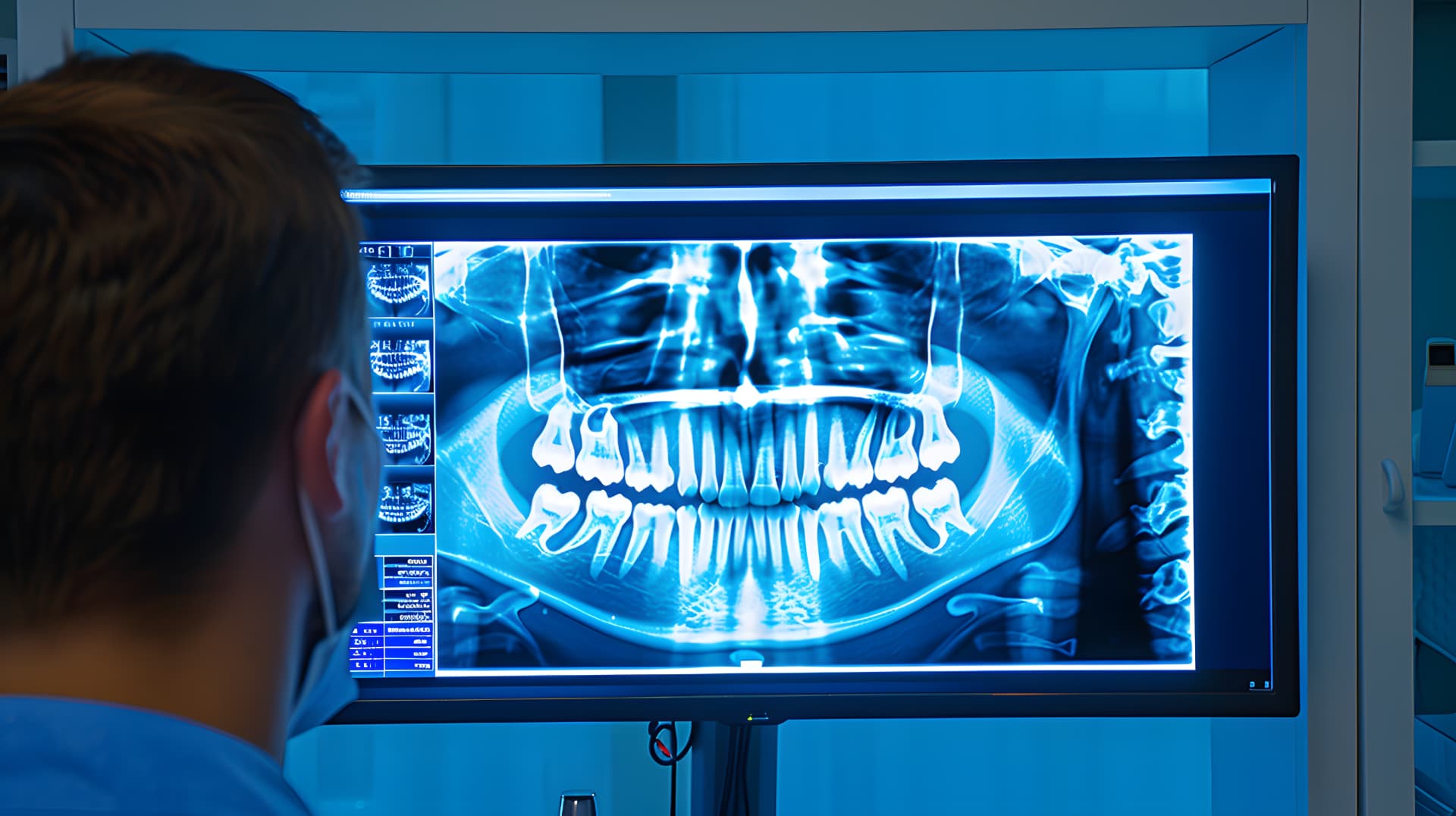
Clinically validated
Optima Stroke
Automated detection of Carotid Artery Calcification from routine panoramic dental X-rays. Published in PLOS Digital Health (2023).

AI Vascular screening
Optima uses advanced AI to reveal vascular risk in the routine medical images clinicians already take, with no new hardware and no disruption to clinical workflows.
Scroll to see a scan ↓
The problem
Billions of medical images are performed every year, yet no systemic vascular screening is in place. Information about arterial health sits unread in images taken for other reasons.

The cost of missing it is severe:
USD 890B+
Global annual cost of stroke, projected to double by 2050.
World Stroke Organization, Global Stroke Fact Sheet 2025 (January 2025).
USD 1.49T
Projected annual US healthcare costs for cardiovascular conditions by 2050, up from USD 393 billion in 2020.
AHA Journals, Forecasting the Economic Burden of Cardiovascular Disease and Stroke in the United States Through 2050 (June 2024).
Up to USD 2.2T
Potential yearly savings for the US health care system by 2040 from investment in prevention, early detection and other proactive measures.
Deloitte, Safeguarding Medicare: Proactive care could unlock $500B in annual program savings (September 2025).
Our approach
Optima is building a new diagnostic layer for global healthcare. It transforms routine medical images into a scalable vascular screening platform, using existing imaging infrastructure. No new hardware. No workflow disruption. A clear path to global scale.
Optima is not a generic AI imaging tool. It is a proprietary diagnostic layer built on unique dataset aggregation and proprietary feature extraction methods for vascular silhouette detection, going beyond visible calcification.
How it works
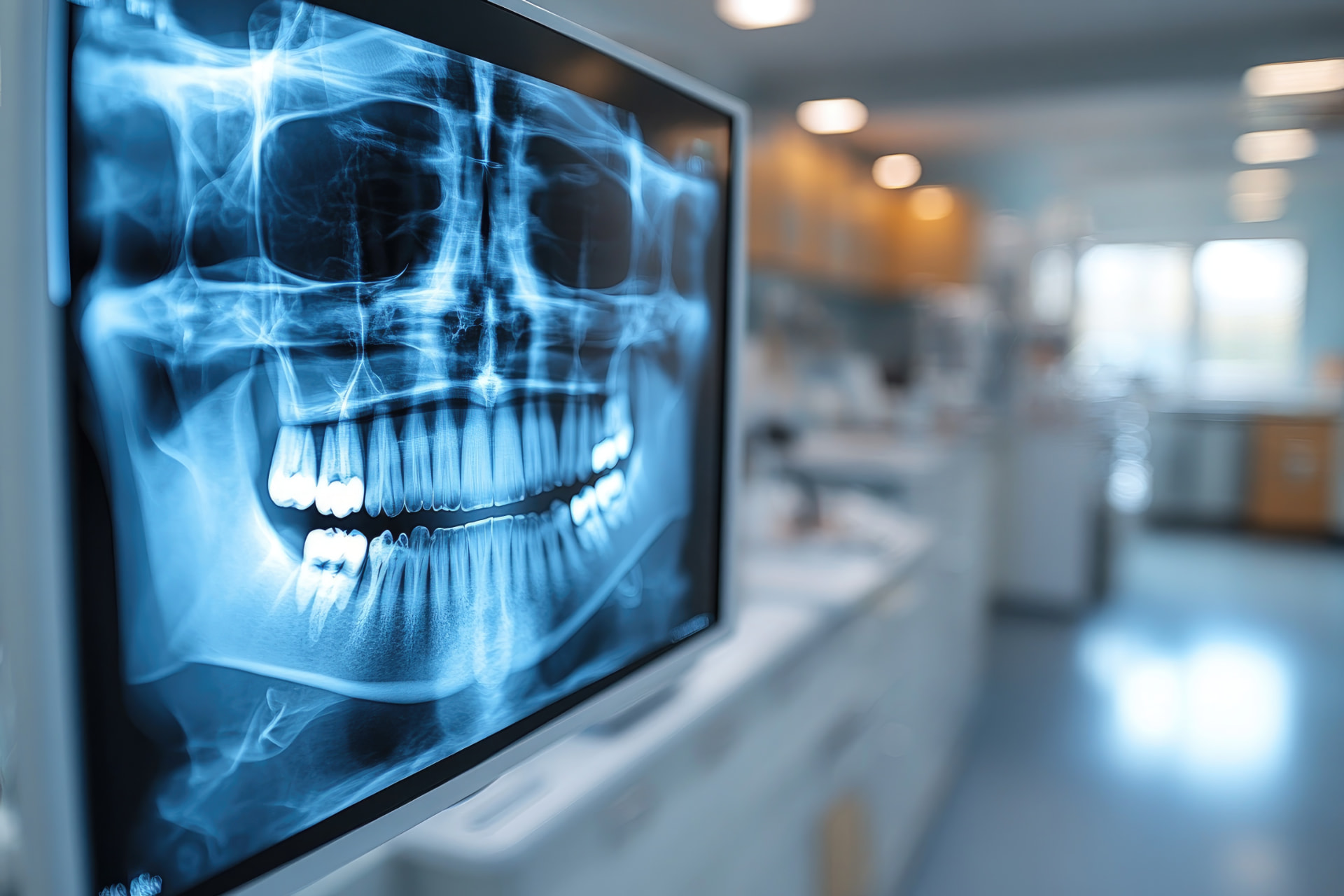
A routine medical image, such as this panoramic dental X-ray, is taken for its usual clinical purpose.
Proprietary neural networks analyze the vascular silhouette in the image.
An automated alert reaches clinicians through existing PACS and EHR systems, so they can consider timely preventive care.
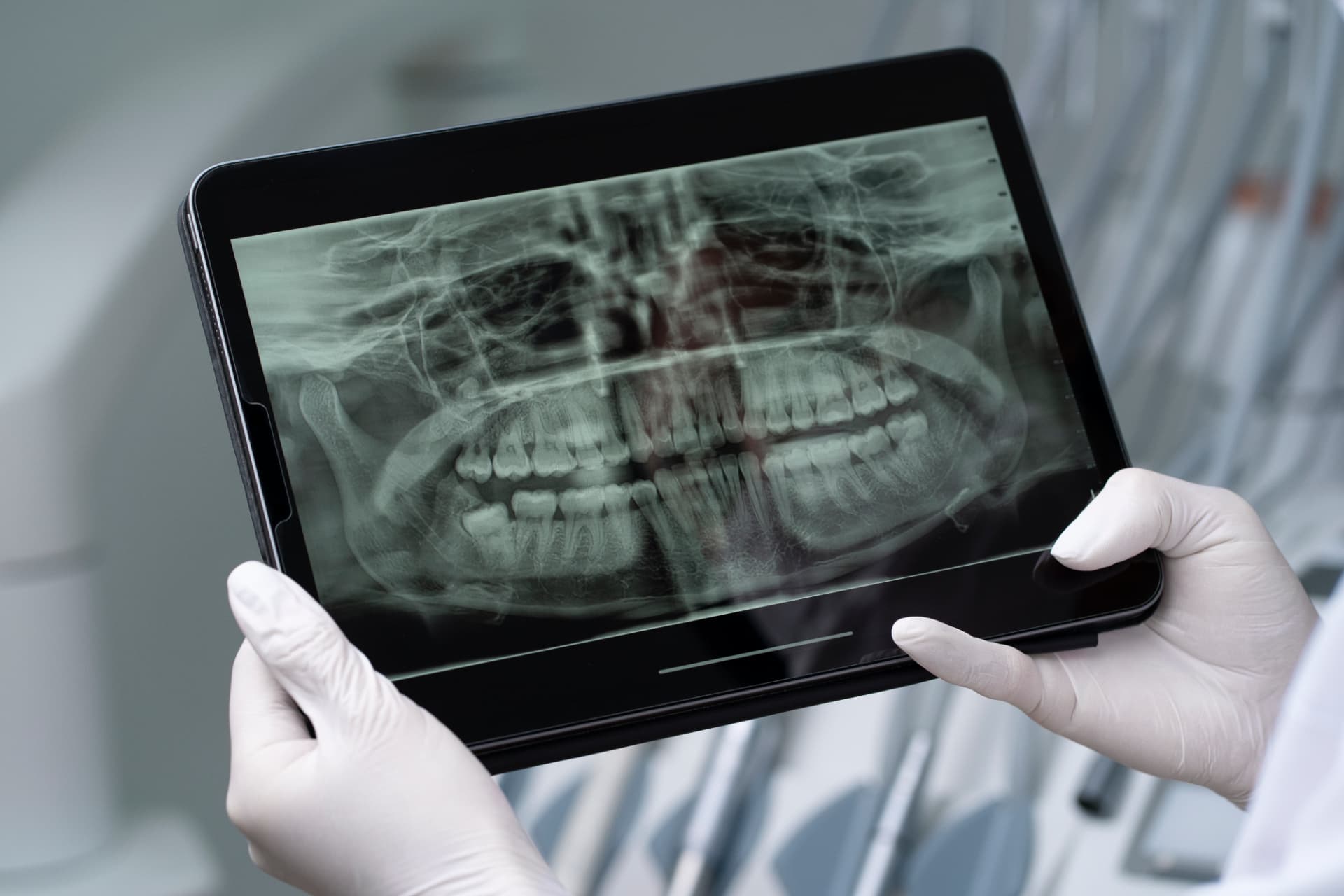
Alert · High Risk
Significant CAC flagged
Sent to clinician via PACS / EHR
Our products
Automated detection of Carotid Artery Calcification from routine panoramic dental X-rays. Published in PLOS Digital Health (2023).
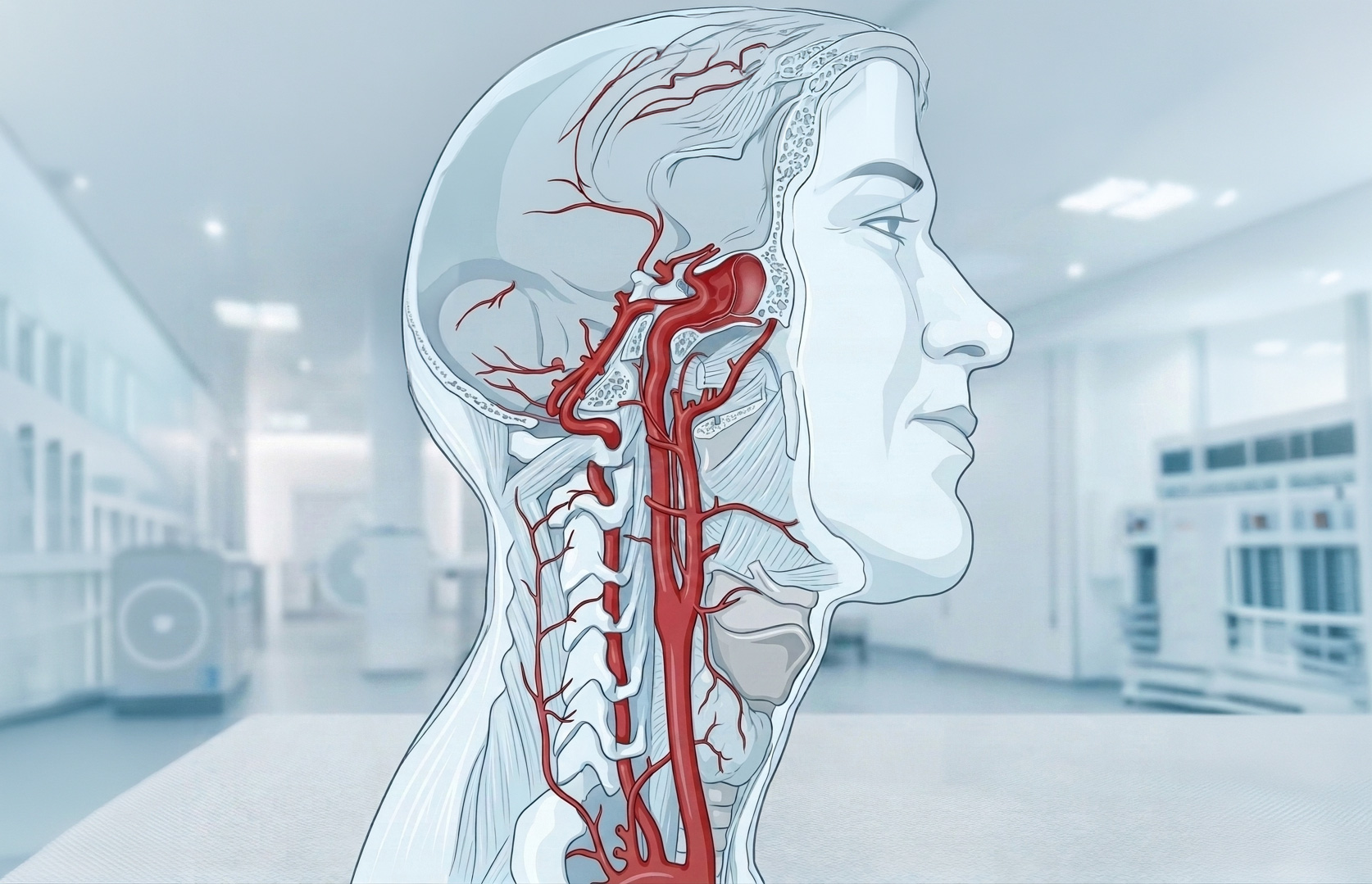
Non-contrast vascular mapping designed to extend the platform across multiple anatomical regions, beginning with radiographs and with MRI envisaged for the next stage. A clinical validation study is being prepared.
Clinical evidence
Optima's first technology has been published in a peer-reviewed journal. Our roadmap is clinical-first: grounded in medical necessity and benchmarked against gold-standard clinical practice.
Optima is moving beyond retrospective research toward population-level opportunistic screening, as a cost-saving infrastructure layer for the future of preventive medicine.
Contact Us
